Medical Ultrasound Imaging
Saturday, 27 April 2024
Info Sheets Out- side     | 'Spatial Resolution' Searchterm 'Spatial Resolution' found in 15 articles 1 term [ • ] - 9 definitions [• ] - 5 booleans [• ]Result Pages : • Spatial Resolution
The spatial resolution describes the smallest distance between two points in the object that can be distinguished as separate details in the image, generally indicated as a length or a number of black and white line pairs per mm (lp/mm). In ultrasound imaging, the spatial resolution means how closely two reflecting tissues - or scattering regions, can be to one another while they can be identified as different reflectors.
•
Standard scanners allow visualizing microbubbles on conventional gray scale imaging in large vascular spaces. In the periphery, more sensitive techniques such as Doppler or non-linear gray scale modes must be used because of the dilution of the microbubbles in the blood pool. Harmonic power Doppler (HPD) is one of the most sensitive techniques for detecting ultrasound contrast agents. Commonly microbubbles are encapsulated or otherwise stabilized to prolong their lifetime after injection. These bubbles can be altered by exposure to ultrasound pulses. Depending on the contrast agent and the insonating pulse, the changes include deformation or breakage of the encapsulating or stabilizing material, generation of free gas bubbles, reshaping or resizing of gas volumes. High acoustic pressure amplitudes and long pulses increase the changes. However, safety considerations limit the pressure amplitude and long pulses decrease spatial resolution. In addition, lowering the pulse frequency increases destruction of contrast bubbles. However, at low insonation power levels, contrast agent particles resist insonation without detectable changes. Newer agents are more reflective and will usually allow gray scale imaging to be used with the advantages of better spatial resolution, fewer artifacts and faster frame rates. Feasible imaging methods with advantages in specific acoustic microbubble properties: Resonating microbubbles emit harmonic signals at double their resonance frequency. If a scanner is modified to select only these harmonic signals, this non-linear mode produces a clear image or trace. The effect depends on the fact that it is easier to expand a bubble than to compress it so that it responds asymmetrically to a symmetrical ultrasound wave. A special array design allows to perform third or fourth harmonic imaging. This probe type is called a dual frequency phased array transducer. See also Bubble Specific Imaging. • 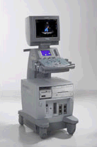 From Siemens Medical Systems;
We see a way to combine quality, versatility, and affordability in a slim, 20-inch wide, ergonomically designed ultrasound system. The new ACUSON CV70™ cardiovascular ultrasound system provides dedicated and complete cardiovascular capabilities in a powerful, all-digital ultrasound system offering exceptional image quality, Doppler sensitivity, color flow sensitivity and spatial resolution − everything you associate with the ACUSON family of cardiovascular products. Specifications for this system will be available soon. •
Breast ultrasound (sonography or ultrasonography) it is an important tool in the characterization of breast lesions, detected with mammography or clinical breast examination. However, a breast sonogram is not approved by the U.S. Food and Drug Administration (FDA) as a screening tool for breast cancer and is used additional to a mammogram. Ultrasound is useful in guiding needles for fine needle aspiration and core biopsies. Breast ultrasound has optimal contrast resolution, but it lacks the spatial resolution of conventional mammography and cannot provide as much detail as a mammogram image. In addition, ultrasound is unable to show tiny calcium deposits (microcalcifications) that are often early indications of breast cancer. See also Biopsy, Interventional Ultrasound, Ultrasound Safety, Side Effect and Ultrasound Regulations. •
Bubble specific imaging methods rely usually on non-linear imaging modes. These contrast imaging techniques are designed to suppress the echo from tissue in relation to that from a microbubble contrast agent. Stimulated acoustic emission (SAE) and phase / pulse inversion imaging mode (PIM) are bubble specific modes, which can image the tissue specific phase. In SAE mode bubble rupture is seen as a transient bright signal in B-mode and as a characteristic mosaic-like effect in velocity 2D color Doppler. PIM are Doppler modes and detect non-linear echoes from microbubbles. In pulse inversion imaging modes the transducer bandwidth extends, resulting in improved spatial resolution and more contrast. See also Contrast Pulse Sequencing, Microbubble Scanner Modification, Narrow Bandwidth, Contrast Medium, Dead Zone. Further Reading: Basics:
Result Pages : | Share This Page Look Ups |
Medical-Ultrasound-Imaging.com
former US-TIP.com
Member of SoftWays' Medical Imaging Group - MR-TIP • Radiology TIP • Medical-Ultrasound-Imaging
Copyright © 2008 - 2024 SoftWays. All rights reserved.
Terms of Use | Privacy Policy | Advertise With Us
former US-TIP.com
Member of SoftWays' Medical Imaging Group - MR-TIP • Radiology TIP • Medical-Ultrasound-Imaging
Copyright © 2008 - 2024 SoftWays. All rights reserved.
Terms of Use | Privacy Policy | Advertise With Us
[last update: 2023-11-06 01:42:00]